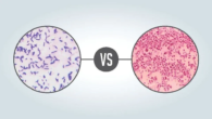


Key Differences Between Mitosis and Meiosis, with a Chart and a Venn Diagram
The Beginning
Cells can either copy themselves to make more cells, or they can get bigger. Human tissues like skin and blood have cells that are constantly dividing. Other tissues, like fat, have cells that are expanding, which is good if you need energy for winter but bad if you want to fit into your favourite jeans. Some cells, like neurons, will never split again after they have reached the end of their differentiation process. These cells are called post-mitotic.
Cells have another choice to make when they copy themselves: do they want to make an exact copy and end up with two cells? Or do they want to make four “half-copies” so that they can reproduce sexually, where fertilisation will fuse their genes back together? You can choose between meiosis and mitosis.
Meiosis and mitosis
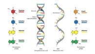
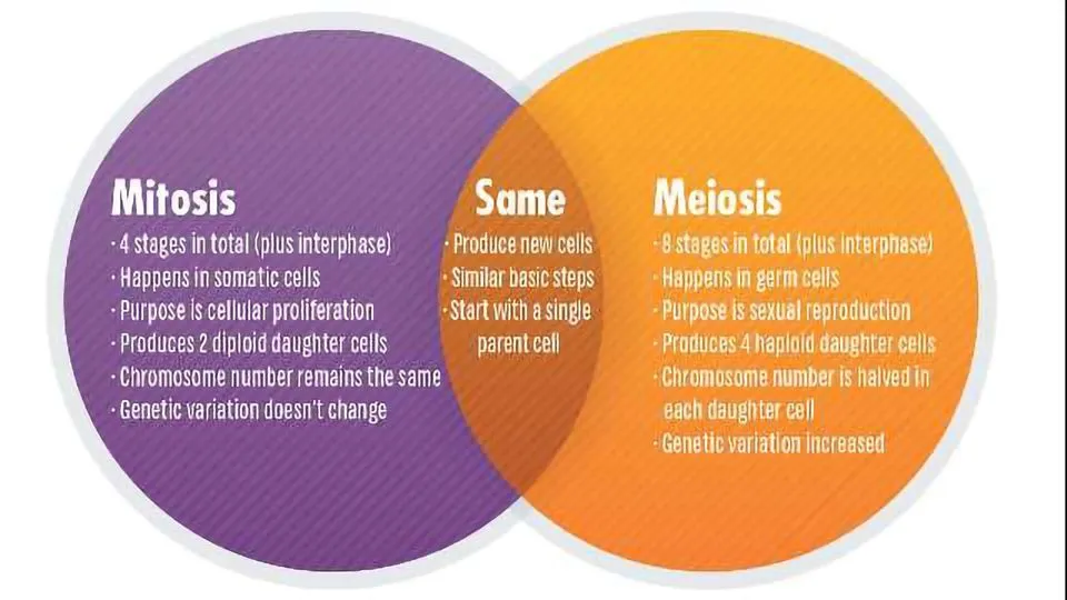
Mitosis and meiosis are the two ways that cells divide. Mitosis is the process by which cells in the body split and make copies of themselves so that the body can grow and heal. Meiosis is the process by which egg and sperm cells are made. During this process, the new cells have half of the genetic material of the parent cell.
Mitosis and Meiosis Are Not the Same
There are some things that are very similar and some very different about these two types of cell division that will be looked at in this piece. The study of these processes and how cell division might go wrong to cause diseases like Down’s Syndrome and cancer will also be talked about.
What is the difference between mitosis and meiosis?
| Mitosis | Meiosis |
| What is the purpose of this process? | |
| In a unicellular organism, the purpose of mitosis is to proliferate as a species. In a multicellular organism, the purpose can be to grow during development, or to repair or regenerate a damaged tissue, for example. | To create gametes with only one copy of the organism’s genetic information, in preparation for sexual reproduction. Various steps in meiosis create opportunity for genetic diversity in the daughter cells. This is the raw substrate for evolution. |
| What is the outcome of this process? | |
| Two diploid cells with identical genetic information. | Four haploid cells with different genetic information. |
| Which organisms perform this process? | |
| Mitosis is performed by unicellular and multicellular eukaryotes. Bacteria have their own version of mitosis called “binary fission”. This is distinct from meiosis as bacteria typically have one circular chromosome, which is not contained within a nucleus, like eukaryotic chromosomes. | Only organisms which perform sexual reproduction. Archaea and bacteria do not do this, so it might be tempting to think that unicellular organisms do not sexually reproduce. However, there are exceptions; budding yeast will form haploid spores under nutritional deprivation. |
| How long does this process take? | |
| Mitosis is usually shorter than meiosis. The process can take over 10 hours for mammalian cells in culture [2], budding yeast can take ~80 minutes to complete a cell cycle [3], whilst bacteria can divide every 20 minutes. | Meiosis has various timescales in different organisms, which can be affected by several factors including temperature and environment of the organism, and the amount of nuclear DNA. The process lasts 6 hours in yeast but can last more than 40 years in human females, due to a developmental hold at prophase I, until ovulation. Other examples are 1-2 days in male fruit flies and ~ 24 days in human males. [1] |
| What is an example of a disease caused by an error in this process? | |
| Uncontrolled mitosis occurs in cancer, where either genes that stop cell division (tumor suppressors) are switched off, or genes that encourage cell division (oncogenes) are overactive. | Errors in meiosis can lead to the wrong number of chromosomes ending up in germ cells, this is called aneuploidy. This can trigger miscarriage, but is occasionally tolerated. One example is Down’s syndrome, caused by trisomy 21. Another example is Klinefelter syndrome, where XY males have an additional X chromosome. |
| Etymology? | |
| Mitosis is the Greek word for thread, after the thread-like chromosomes that can be seen under the microscope in dye-stained cells during cell division. | Meiosis means a “lessening” in Greek. This refers to the outcome of meiosis, where the genetic information in each new cell is halved. |
| First described by? | |
| Walther Flemming in his 1882 work “Cell substance, nucleus and cell division.” [5] | Oskar Hertwig described the fusion of egg and sperm in the transparent sea urchin egg in 1876. [4] |
How to Set the Scene for Mitosis vs. Mitosis
A part of the “cell cycle” is cell division. Cells follow their own schedules, just like your day does from morning to night. There are four key stages in the cell cycle, which are called G1, S phase, G2, and mitosis (or meiosis). Cells can also take a break from the cell cycle by going into a state called G0 or senescence. Some cells stay in G0 all the time. Outside growth factors, like Nerve Growth Factor (NGF), which helps neurons grow, can encourage cells in G1 or G0 to move on to the next stage of the cycle. There is a special “point of no return” in G1 called the restriction point. This is where cells stop responding to growth factors and will keep moving to S phase no matter what. There are also signals inside the cell that tell it to move forward. These are proteins called cyclins, and cyclin B is the cyclin that speeds up mitosis. The S phase is very important because this is when the cell’s whole genome is copied using a method called semi-conservative DNA replication.
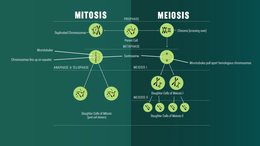
A look at the steps of meiosis and mitosis
Mitosis has five stages: interphase, prophase, metaphase, anaphase, and telophase. Cytokinesis can happen after telophase. This word “interphase” refers to all the steps before mitosis, which are the G1, S, and G2 phases. Anaphase I, telophase I, cytokinesis I, prophase II, metaphase II, anaphase II, telophase II, and finally cytokinesis II make up the stages of meiosis. Read our full reasoning below:
| Summary | |
| Meiosis and mitosis both have a prophase, metaphase, anaphase, telophase and cytokinesis. | |
| In meiosis, prophase, metaphase, anaphase and telophase occur twice. The first round of division is special, but the second round is more like mitosis. | In mitosis, prophase, metaphase, anaphase and telophase occur once. |
| Prophase | |
| Chromosomes condense and the centrosomes begin to form an early spindle. | |
| Meiotic prophase I is much longer that mitotic prophase.During prophase I homologous chromosomes make contacts with each other called chiasmata and “crossing over” occurs. This is where chromosomes exchange sections of DNA. This is important for generating genetic diversity but is also crucial mechanically to hold homologous chromosomes together. | Mitotic prophase is much shorter that meiotic prophase I.There is no crossing over in mitosis. |
| Metaphase | |
| In metaphase II of meiosis, and metaphase of mitosis, chromosomes line up along the metaphase plate due to the action of microtubule spindle fibers emanating from the centrosomes located at opposite cell poles. These fibers are attached to the chromosomes by kinetochores at the centromeres of the chromosomes. | |
| In meiotic metaphase I pairs of homologous chromosomes line up along the metaphase plate.The way in which the homologous pairs are oriented randomly with respect to the cell poles is referred to as the law of independent assortment and ensures a random and independent distribution of chromosomes to the daughter cells of meiosis I and ultimately to the haploid gametes at the end of meiosis II. | In mitotic metaphase a single chromosome/ pair of chromatids line up along the metaphase plate.Sister chromatids are identical and so the orientation of the chromosome doesn’t carry any meaning. |
| Anaphase | |
| In anaphase, chromosomes are split to opposite poles of the cell. | |
| In anaphase of meiosis I cohesin at the centromeres of the chromosomes is not cleaved and it therefore continues to hold sister chromatids together as the homologous chromosomes are segregated to opposite cell poles. | In anaphase of mitosis (and meiosis II), cohesin protein holding the centromeres of the sister chromatids together is cleaved, allowing the sister chromatids to segregate to opposite poles of the cell, at which point they are called chromosomes. |
| Telophase | |
| A nuclear membrane reforms around the newly separated chromosomes, which begin to uncoil, becoming less condense. The spindle microtubules disassociate. Each daughter cell will inherit one centrosome. | |
| Cytokinesis | |
| The cell plasma membrane pinches, to leave two daughter cells with separate plasma membranes. | |
| In meiosis, cytokinesis must occur twice: once after telophase I and again, after telophase II. | In mitosis, cytokinesis does not always occur, some cells divide and are multinucleate, like muscle cells. |
How to Improve Your Memory
You can also think about what is happening to the chromosomes, centrosomes, nuclear membrane, and cell plasma membrane at each stage of the process to better understand how it moves forward. You can see how to do this for mitosis here. Why not try to make this table for meiosis too?
Another useful tool is the memory “I Prefer Mating At Teatime” by Chamillionaire, which can help you remember the order of the steps in mitosis.
| Mitosis Stage | Chromosomes |
| Interphase | Are uncondensed but are still organized. The entire genome is replicated to create two identical semi-conserved copies of each chromosome. |
| Prophase | Condense. Duplicated chromosomes are called sister chromatids. |
| Metaphase | Align along the metaphase plate, the midpoint between the two centrosomes. Sister chromatids are joined at the centromere by proteins that form a structure called a kinetochore. |
| Anaphase | Cohesin is cleaved at the centromere of chromosomes, resulting in sister chromatids being pulled to opposite poles of the cell. |
| Telophase | Chromosomes begin to uncoil, becoming less condensed. |
| Cytokinesis | Chromosomes have returned to their interphase structure. This is a topic of much research, but it seems as though each chromosome occupies its own territory within the nucleus. |
| Mitosis Stage | Centrosomes |
| Interphase | The centrosome is duplicated. |
| Prophase | Microtubules begin to form an early mitotic spindle between the duplicated centrosomes. |
| Metaphase | The two centrosomes are now located at opposite poles of the cell. |
| Anaphase | Microtubules emanating from the centrosomes shrink as the tension holding the chromosomes at the metaphase plate is broken by cohesin cleavage. |
| Telophase | The centrosomes remain segregated to opposite sides of the cell. Each daughter cell will receive one centrosome comprised of two centrioles. |
| Cytokinesis | Centrosomes signal to the cell that it is okay to proceed with cytokinesis. Research shows that cells where centrosomes are destroyed with a laser beam cannot undergo cytokinesis. |
| Mitosis Stage | Nuclear Membrane |
| Interphase | Intact. |
| Prophase | Intact. |
| Metaphase | In higher eukaryotes like vertebrates, by the time metaphase occurs the nuclear envelope has broken down. This is caused by phosphorylation of nuclear lamin proteins. |
| Anaphase | Broken down. |
| Telophase | A nuclear envelope reforms around the chromosomes in each daughter cell. |
| Cytokinesis | Intact. |
| Mitosis Stage | Plasma Membrane |
| Interphase | Intact. |
| Prophase | Intact. |
| Metaphase | Intact. |
| Anaphase | Intact. |
| Telophase | Intact. |
| Cytokinesis | Pinches to form two separate membranes around the two daughter cells. |
Myosis vs. Mitosis The Venn Diagram
Questions for active study
Scientists have been interested in the complex dance of molecular machinery that happens when cells divide for hundreds of years. Microscopy has come a long way since its early days of looking at metaphase chromosomes under a light microscope. Today, there are more advanced tools that can answer problems at the molecular level. There have also been many awards for research into the cell cycle. For example, Tim Hunt, Paul Nurse, and Leland Hartwell won the Nobel Prize in Physiology or Medicine in 2001 for finding cyclins and cyclin-dependent kinases, which are the main proteins that control the cell cycle [6]. But, even though we’ve made progress, there are still a lot of questions.
How do cells make sure that chromosomes are properly separated during mitosis?
There is just one way for mitosis to go right, but many ways for it to go wrong. For instance, if microtubules and chromosomes don’t touch correctly in the early stages of mitosis, chromosomes can become out of place, which can cause sister chromatids to separate in the wrong way. How can the cell be sure that it is time to do cytokinesis when mitosis is almost over? The chromosome passenger complex (CPC) is like a molecular guardian angel that keeps the process honest at many points during mitosis. The CPC is found all over the chromosomes at the start of mitosis and changes the chromatin. During mitosis, it goes to the centromeres of the chromosomes to stop microtubules from attaching incorrectly, and just before cytokinesis, it gets to the central spindle. So, one question that scientists are still looking into is how the CPC so gracefully moves around during mitosis to save the day.
- Vader, G., Medema, R. H., & Lens, S. M. (2006). Aurora-B is being led through mitosis by the chromosome passenger complex. It’s in The Journal of cell biology (173(6), 833–837).
•Zou, L., Kabeche, L., Nguyen, H. D., & Buisson, R. (2018). ATR pathway that is special to mitosis and driven by R loops helps chromosomes separate correctly. It’s in Science, 359(6371), 108–114.
How are identical chromosomes held together during meiosis I and then split apart?
In metaphase of mitosis and metaphase II of meiosis, the protein cohesin keeps sister chromatids together. You may remember this from earlier. In meiosis I, however, homologous chromosomes need to be held together during metaphase I. During anaphase I, these bonds are quickly broken. This is possible because of an amazing cellular zipper known as the synaptonemal complex (SC). This zipper needs to be strong enough to hold chromosomes together, but it also needs to be able to be taken apart just as easily. If it can’t do both, homologous chromosomes won’t separate correctly in anaphase I, which could cause genetic difference in the daughter cells that could be very bad. A lot of people are interested in figuring out how this zip comes apart.